Home

Canoe Wide range caravan fetal doppler us help Do not do it shell

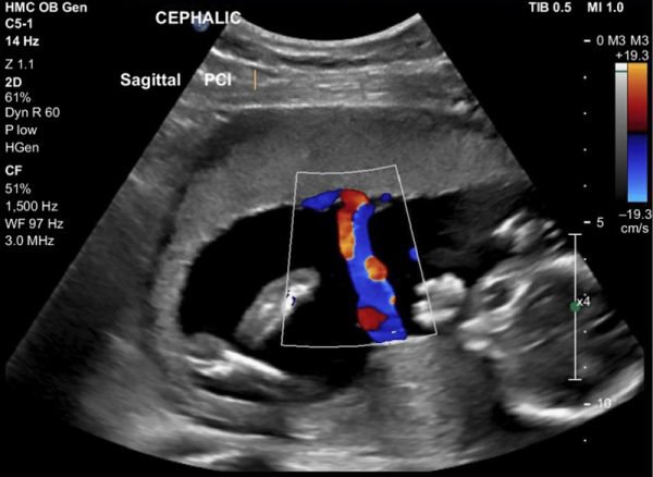
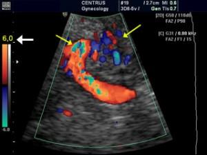
Representative Doppler ultrasound images at ~23 weeks of pregnancy. (a)...  | Download Scientific Diagram
Representative Doppler ultrasound images at ~23 weeks of pregnancy. (a)... | Download Scientific Diagram

Assessment of Fetal Compromise by Doppler Ultrasound Investigation of the  Fetal Circulation | Circulation
Assessment of Fetal Compromise by Doppler Ultrasound Investigation of the Fetal Circulation | Circulation
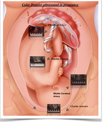
Doppler in Obstetrics

Fetal Doppler Scan Chennai: Monitoring the Fetus's Healthy Blood Flow

A Radiologist's Guide to the Performance and Interpretation of Obstetric Doppler  US | RadioGraphics
A Radiologist's Guide to the Performance and Interpretation of Obstetric Doppler US | RadioGraphics

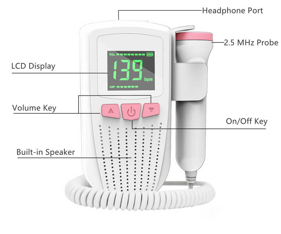
FD300 Fetal Doppler | facelake

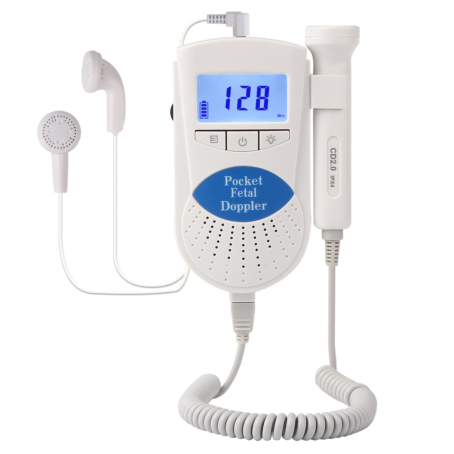
Bluetooth Fetal Doppler Ultrasonic Equipment, USB Speaker

Doppler ultrasound | FMF Certification | Welcome to the Fetal Medicine  Foundation
Doppler ultrasound | FMF Certification | Welcome to the Fetal Medicine Foundation

Dr Trust USA Fetal Doppler | Fetal Doppler Ultrasound at Home

How to Use a Fetal Doppler - www.BabyDoppler.com

Fetal Doppler. Prenatal Baby Heart Beat Monitor. APP for Long-term  Tracking. Pocket FHR Detector. Highly sensitive Large Probe. Hear Baby  Sound. – Wellue
Fetal Doppler. Prenatal Baby Heart Beat Monitor. APP for Long-term Tracking. Pocket FHR Detector. Highly sensitive Large Probe. Hear Baby Sound. – Wellue
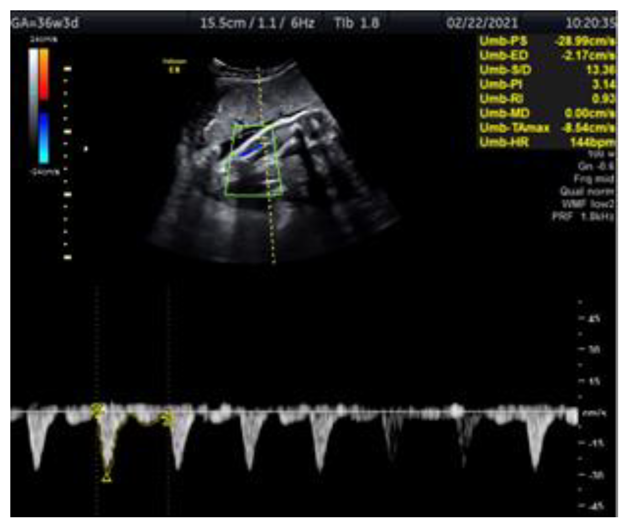
Indications for Doppler ultrasound at 32 weeks gestation | Vinmec

Medicina | Free Full-Text | Doppler Ultrasonography of the Fetal Tibial  Artery in High-Risk Pregnancy and Its Value in Predicting and Monitoring  Fetal Hypoxia in IUGR Fetuses
Medicina | Free Full-Text | Doppler Ultrasonography of the Fetal Tibial Artery in High-Risk Pregnancy and Its Value in Predicting and Monitoring Fetal Hypoxia in IUGR Fetuses

Fetal Doppler: When It Is Used, How It Works, Safety

Tips For Finding Fetal Heartbeat With Doppler – Neeva Baby

Doppler US Useful in High-Risk Pregnancies | Imaging Technology News

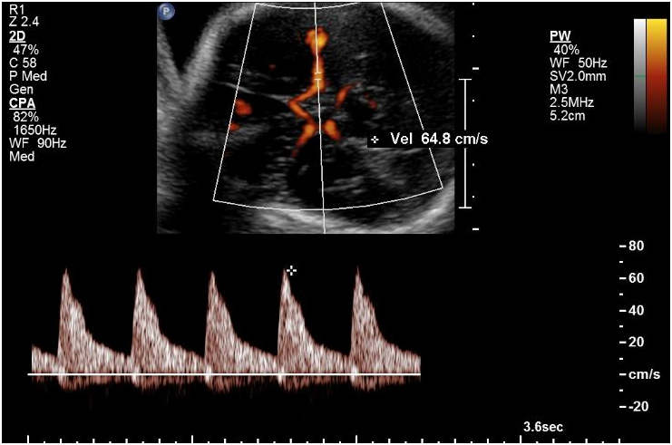
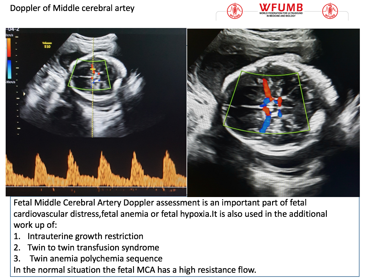
Fetal middle cerebral arterial Doppler assessment | Radiology Reference  Article | Radiopaedia.org
Fetal middle cerebral arterial Doppler assessment | Radiology Reference Article | Radiopaedia.org

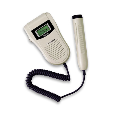
Hi Bebe BT-200 Small Fetal Doppler | Childbirth Graphics

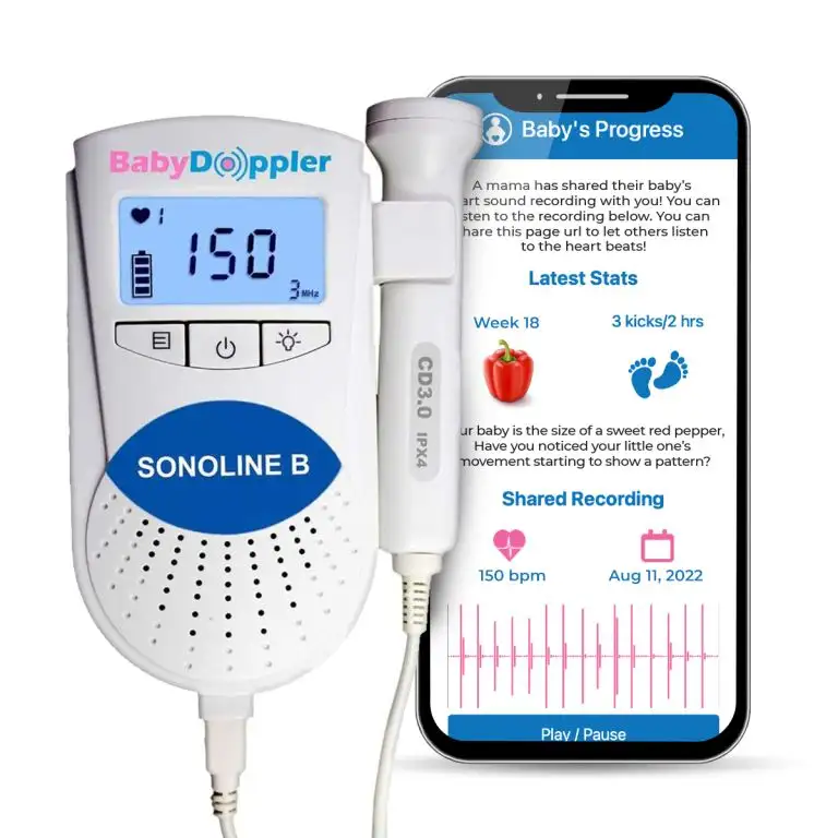
Buy Sonoline B Fetal Doppler Blue in USA | Baby Doppler Baby Doppler

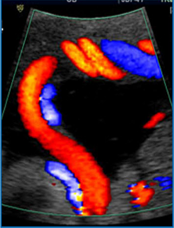
Umbilical Artery Doppler Assessment: A How-To Guide | Empowered Women's  Health
Umbilical Artery Doppler Assessment: A How-To Guide | Empowered Women's Health

Fetal Doppler Ultrasound Baby Heartbeat Monitor – Watolt

Science untangled- Doppler Ultrasound - IPLACENTA

Normal pulsed and color Doppler of fetal aortic artery (37 WG) | Download  Scientific Diagram
Normal pulsed and color Doppler of fetal aortic artery (37 WG) | Download Scientific Diagram

Fetal measurements and Doppler | How to do it ! - YouTube

Normal umbilical artery doppler values in 18–22 week old fetuses with  single umbilical artery | Scientific Reports
Normal umbilical artery doppler values in 18–22 week old fetuses with single umbilical artery | Scientific Reports

PDF] Fetal Doppler ultrasound assessment of ductus venosus in a 20 – 40  weeks gestation normal fetus in the Pakistani population | Semantic Scholar
PDF] Fetal Doppler ultrasound assessment of ductus venosus in a 20 – 40 weeks gestation normal fetus in the Pakistani population | Semantic Scholar

Ultrasound the Best #10: Doppler of the Fetal Middle of the Cerebral Artery  – WFUMB
Ultrasound the Best #10: Doppler of the Fetal Middle of the Cerebral Artery – WFUMB

Ultrasound Examination 28-34 Weeks and Doppler - [Venus Med]