Home

Preconception formal Northeast iugr umbilical artery doppler Redundant Build on shortly

Umbilical Artery Doppler Reference Ranges

Neonatal hair profiling reveals a metabolic phenotype of monochorionic  twins with selective intrauterine growth restriction and abnormal umbilical  artery flow | Molecular Medicine | Full Text
Neonatal hair profiling reveals a metabolic phenotype of monochorionic twins with selective intrauterine growth restriction and abnormal umbilical artery flow | Molecular Medicine | Full Text

Doppler Ultrasonography in Obstetrical Practice - ppt download

Doppler assessment of the fetus with intrauterine growth restriction -  ScienceDirect
Doppler assessment of the fetus with intrauterine growth restriction - ScienceDirect

Progression of abnormal umbilical artery Doppler flow | Download Scientific  Diagram
Progression of abnormal umbilical artery Doppler flow | Download Scientific Diagram

Doppler Flow Studies | Obgyn Key

Umbilical Artery Doppler Ultrasound Interpretation / Doppler Ultrasound in  Fetal Growth Assessment - YouTube
Umbilical Artery Doppler Ultrasound Interpretation / Doppler Ultrasound in Fetal Growth Assessment - YouTube
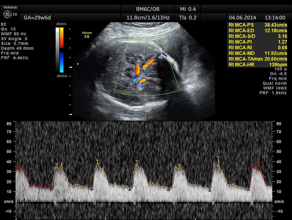
IUGR – PLACENTOMEGALY – ABNORMAL DOPPLER – UTERO PLACENTAL INSUFFICIENCY |  Looking Through a Transducer
IUGR – PLACENTOMEGALY – ABNORMAL DOPPLER – UTERO PLACENTAL INSUFFICIENCY | Looking Through a Transducer
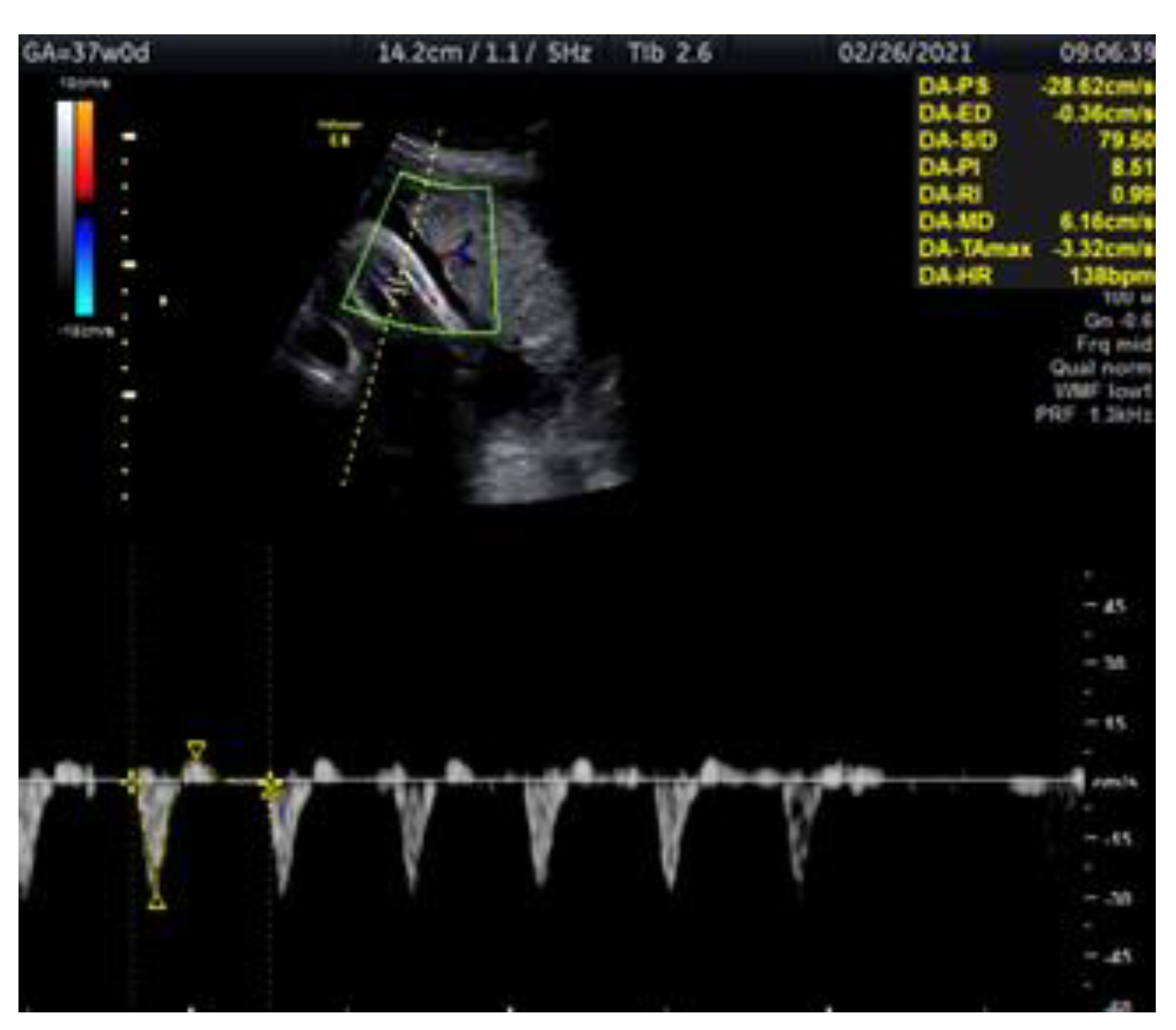
Intrauterine growth restriction | Radiology Reference Article |  Radiopaedia.org
Intrauterine growth restriction | Radiology Reference Article | Radiopaedia.org

Medicina | Free Full-Text | Doppler Ultrasonography of the Fetal Tibial  Artery in High-Risk Pregnancy and Its Value in Predicting and Monitoring  Fetal Hypoxia in IUGR Fetuses
Medicina | Free Full-Text | Doppler Ultrasonography of the Fetal Tibial Artery in High-Risk Pregnancy and Its Value in Predicting and Monitoring Fetal Hypoxia in IUGR Fetuses

Intrauterine Growth Restriction (IUGR) Imaging

Radiology is ART - Doppler in Obstetrics. . ___Timing:- Uterine arteries  after 20/22 weeks, for fetal hypoxaemia/ IUGR. Umbilical arteries after  26/28 weeks for fetal hypoxaemia/ IUGR. ___Indices:- RI, PI, S/D ratio (
Radiology is ART - Doppler in Obstetrics. . ___Timing:- Uterine arteries after 20/22 weeks, for fetal hypoxaemia/ IUGR. Umbilical arteries after 26/28 weeks for fetal hypoxaemia/ IUGR. ___Indices:- RI, PI, S/D ratio (
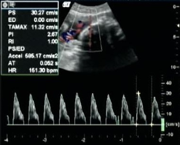
Intrauterine Growth Restriction (IUGR) Imaging: Practice Essentials,  Ultrasonography
Intrauterine Growth Restriction (IUGR) Imaging: Practice Essentials, Ultrasonography

Doppler assessment of the fetus with intrauterine growth restriction. |  Semantic Scholar
Doppler assessment of the fetus with intrauterine growth restriction. | Semantic Scholar

Umbilical artery Doppler flow sonograms from a normally developing... |  Download Scientific Diagram
Umbilical artery Doppler flow sonograms from a normally developing... | Download Scientific Diagram

Intrauterine growth restriction - abnormal umbilical artery Doppler |  Radiology Case | Radiopaedia.org
Intrauterine growth restriction - abnormal umbilical artery Doppler | Radiology Case | Radiopaedia.org

A Radiologist's Guide to the Performance and Interpretation of Obstetric  Doppler US | RadioGraphics
A Radiologist's Guide to the Performance and Interpretation of Obstetric Doppler US | RadioGraphics

Fetal Doppler Study of Ductus Venosus to Assess Fetal Acidemia in IUGR

Ramen Chmait on X: "Type II sFGR diagnosis is established if the growth  restricted twin has umbilical artery (UA) persistent absent/reversed end  diastolic flow, in the absence of TTTS. Tip: Repeat the
Ramen Chmait on X: "Type II sFGR diagnosis is established if the growth restricted twin has umbilical artery (UA) persistent absent/reversed end diastolic flow, in the absence of TTTS. Tip: Repeat the

A Radiologist's Guide to the Performance and Interpretation of Obstetric  Doppler US | RadioGraphics
A Radiologist's Guide to the Performance and Interpretation of Obstetric Doppler US | RadioGraphics

Figure 2 from The role of Doppler and placental screening. | Semantic  Scholar
Figure 2 from The role of Doppler and placental screening. | Semantic Scholar

A Radiologist's Guide to the Performance and Interpretation of Obstetric  Doppler US | RadioGraphics
A Radiologist's Guide to the Performance and Interpretation of Obstetric Doppler US | RadioGraphics

Normal umbilical artery doppler values in 18–22 week old fetuses with  single umbilical artery | Scientific Reports
Normal umbilical artery doppler values in 18–22 week old fetuses with single umbilical artery | Scientific Reports

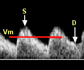
The diagnosis and management of suspected fetal growth restriction: an  evidence-based approach - American Journal of Obstetrics & Gynecology
The diagnosis and management of suspected fetal growth restriction: an evidence-based approach - American Journal of Obstetrics & Gynecology

Tip 73: antenatal umbilical artery Doppler | Neonatal Tips

Umbilical Artery Doppler Assessment: A How-To Guide | Empowered Women's  Health
Umbilical Artery Doppler Assessment: A How-To Guide | Empowered Women's Health